
EMG reads the muscle's own electrical answer.
Every twitch starts as an electrical signal inside the muscle. Electromyography measures that signal at the skin, so the reading doesn't depend on how far a thumb can move, and it keeps working when the arms are tucked out of sight.
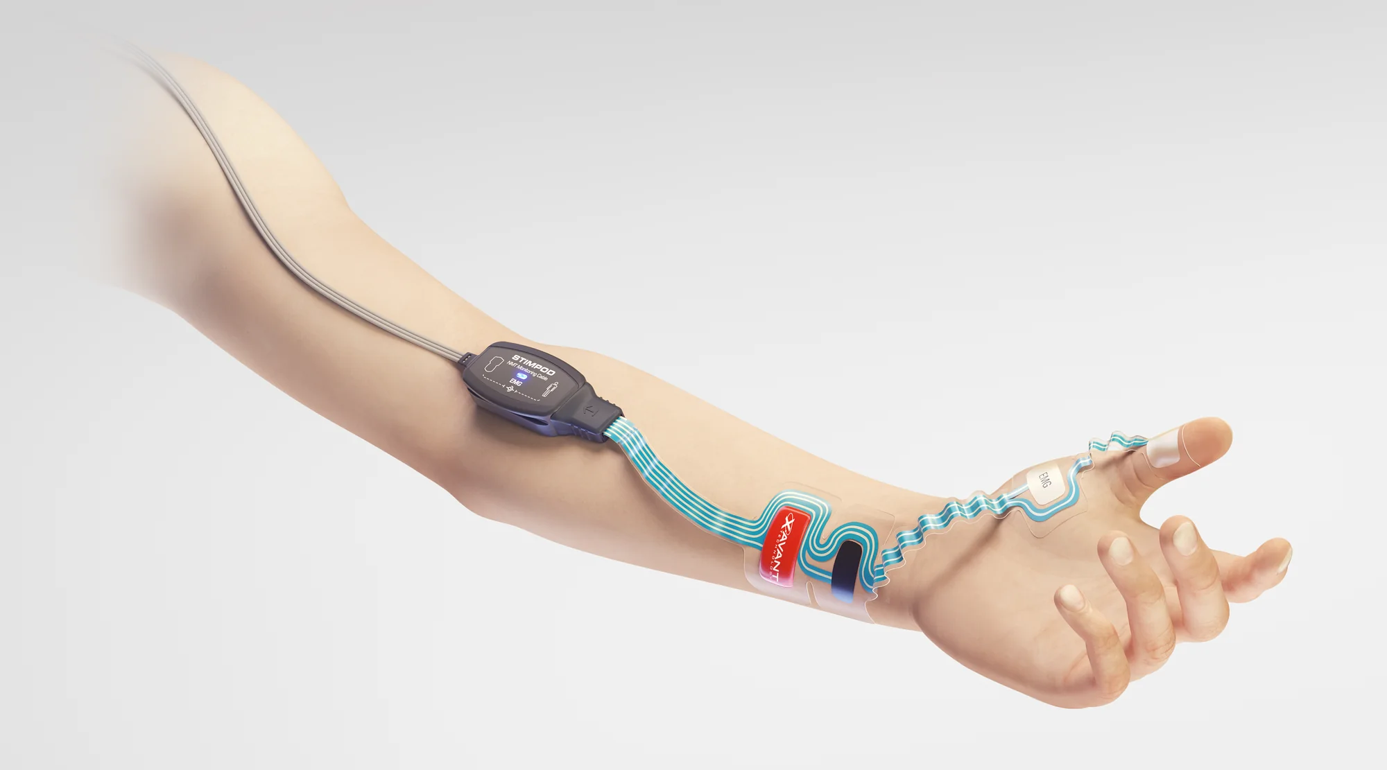
From a pulse at the wrist to a number on the screen.
A train-of-four takes two seconds. Here is what happens in that time, from the first pulse to the TOF ratio you read.
The stimulus
Stimpod™ sends four brief current pulses along the ulnar nerve at the wrist, half a second apart.
The junction
Each pulse reaches the neuromuscular junction, where the blocking drug competes with acetylcholine for the receptors.
The muscle fires
The fibres that are still free to respond fire together. Their combined signal is the compound muscle action potential, the CMAP.
The reading
The recording pad picks up each wave, from the smallest response Stimpod™ counts, 400 µV, up to ±30 mV. The fourth wave over the first is the TOF ratio.
Watch a train‑of‑four being read.
Set the depth of block and fire a train. Each pulse reaches the electrodes first as a sharp stimulus artefact, and the muscle answers a few milliseconds later. The Stimpod™ shows only the muscle's wave, measured by its height and its area, and draws it over the grey reference pulse.
At the electrodes
Four pulses, 0.5 seconds apart- Peak-to-peak heightThe wave's height, from its highest point to its lowest.
- Area under the curveAll the electrical activity in the wave, above and below the line.
- Stimulus artefactThe stimulation pulse itself, picked up a few milliseconds before the muscle answers.
On the Stimpod™ screen
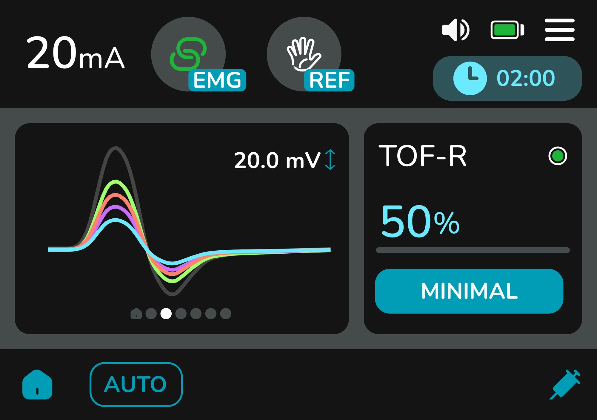
- The grey wave is the reference pulse. It shows the response's height before the block, so you can see how far T1 has dropped.
- The number in the corner is the peak-to-peak height of the latest wave, in millivolts. It updates with every twitch, so after a train it shows the height of T4.
- T1 to T4 arrive in their own colours on the same scale, so the fade from the first wave to the fourth is the TOF ratio.
- The stimulus artefact is not on the screen. You see only the muscle's wave, the part that is measured.
A modelled illustration built for this page. The waves are not recordings, and this is not the Stimpod™ algorithm.
Stimpod™ reads the height and the area of every wave.
Peak-to-peak amplitude measures how tall a wave is, from its highest point to its lowest. Area under the curve measures how much electrical activity the whole wave carries. Most EMG monitors pick one of the two. Stimpod™ uses both to determine each result.
On a clean signal the two tell the same story: a 2024 analysis of 5,559 paired TOF ratios found them clinically interchangeable. They part ways when the signal isn't clean. A stimulus artefact is a tall, narrow spike, so it inflates the height and barely touches the area. A drifting baseline shifts the whole trace, so it swells the area and leaves the height alone.
Epstein et al · BJA Open 2024;11:100293Each feature is blind where the other can see, so a result read from both has two ways to be checked.
Made for the cases where you can't see the hand.
Tucked arms, robots and laparoscopy
EMG reads electricity, not movement. The thumb can be wrapped, strapped to the side or under the drapes, and the reading carries on.
No free thumb, no preload
Nothing has to swing freely or be held in a set position, so there's no hand adapter to fit and nothing for the drapes to press against.
A steadier baseline
Repeated stimuli hardly change the size of the CMAP, so EMG is far less prone to the gradual drift that affects movement-based readings over a long case.
Koo · Anesth Pain Med 2025;20(3):222-229The smallest patients
With the small electrode, the same monitor covers children from 28 days after birth, on either hand.
Tested against the gold standard.
A mechanomyograph measures the actual force of the thumb and is the reference every NMT monitor is judged against. A team at the University of Washington compared the Stimpod™ EMG with one twice, through real blockade in surgery in 2023 and with no blocking drug in 2024.
Across both studies the Stimpod™ EMG read like the mechanomyograph. During blockade its TOF ratios tracked the reference with a correlation of 0.94 and a bias of 0.028, and with no blocking drug its readings held as steady as the reference's, with a spread of 0.021 against 0.022. The authors called its results comparable to the mechanomyograph's in 2023 and similar to it in 2024.
During blockade
2023- TOF ratio415 pairs
- Correlation 0.94, bias 0.028
- Post-tetanic count338 pairs
- Weighted kappa 0.80, substantial agreement
23 patients, 1,088 paired readings, same hand as the mechanomyograph.
The authors concluded“The electromyograph produced results that were comparable to the mechanomyograph.”
Wedemeyer et al · J Clin Monit Comput 2024;38(1):205-212 · online Oct 2023With no blocking drugGauge standard deviation of the TOF ratio, lower is tighter
2024Stimpod™ EMG: 1,130 readings in 7 patients, mean 1.01. Mechanomyograph: 2,376 readings in 23 patients, mean 0.99.
The authors concluded“[Stimpod™ EMG] measured the train-of-four ratio with minimal overshoot and variability, similar to a mechanomyograph.”
Wedemeyer et al · Anesthesiology 2024;141(2):262-271 · Table 2Two electrodes, sized for every patient.
The ripple electrode
A patented, skin-like strip that fits either hand and any hand size. The ripples let it move with the skin, so it stays put through long cases and patient movement.
- 1Cable module: connects the strip to the reusable NMT Monitoring Cable EMG, 1.8 m or 3.5 m.
- 2Stimulating pads, over the ulnar nerve at the wrist.
- 3Recording pad, over the muscles between thumb and index finger.
- 4Reference pad, on the thumb.
Skin preparation, placement steps and warnings are in the Instructions for Use.
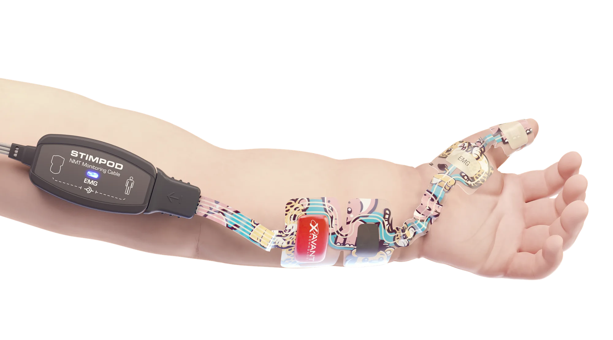
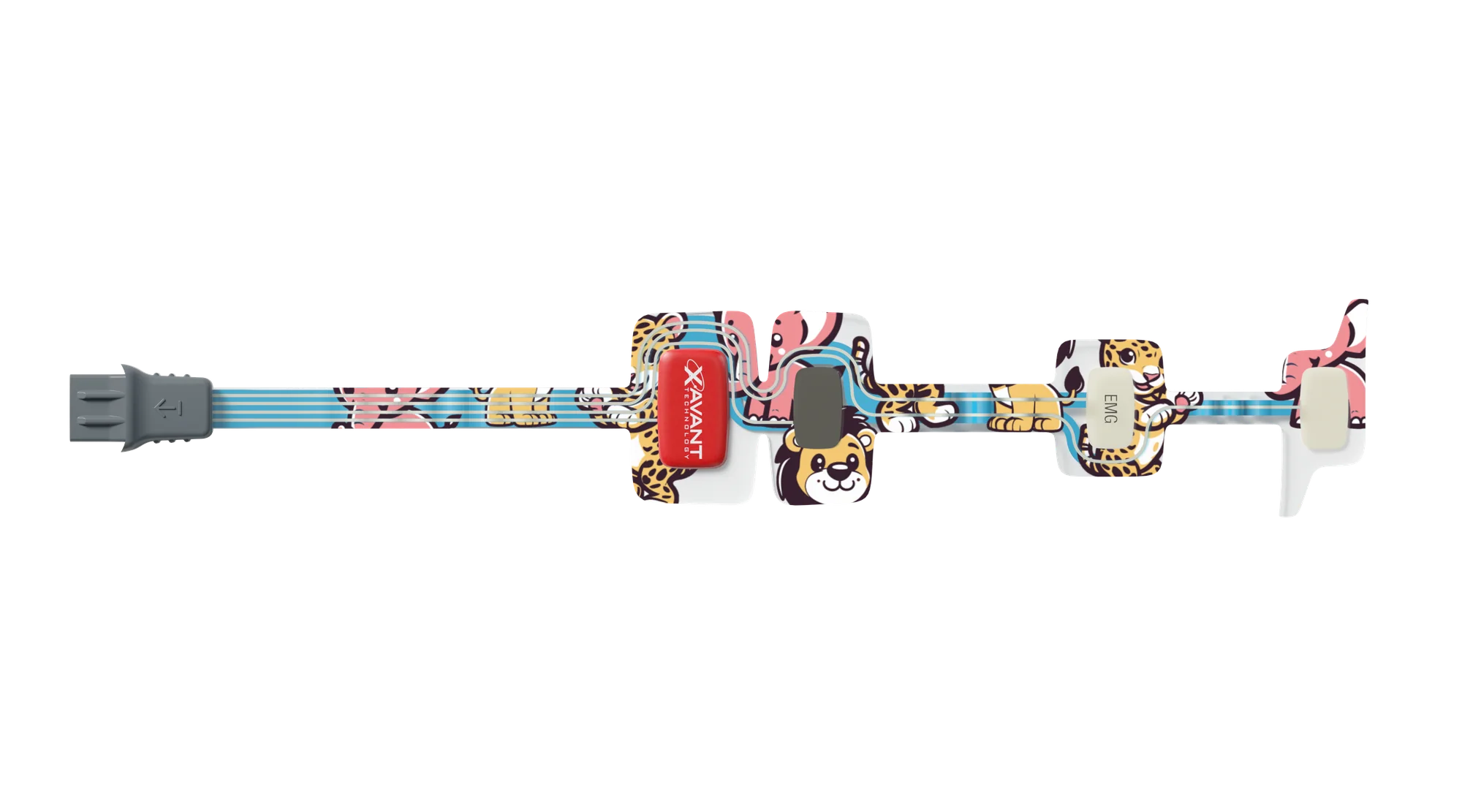
The small electrode
The same EMG monitoring, sized for children from 28 days after birth. It goes on either hand and is approved to stay on for up to a full day.
- Product code
- XT-45009S
- Patients
- Children from 28 days after birth
- Hand
- Left or right
- Adhesion
- Approved for up to 24 hours
- Pack
- Box of 20
- Shelf life
- 24 months
- Works with
- Stimpod™ NMS450X+ and NMS450X, through the NMT Monitoring Cable EMG
See the signal behind the number.
In EMG mode the Stimpod™ draws each response as it arrives, next to the TOF ratio. If a wave looks wrong, you see it before you trust the number.
Explore the NMS450X+ →See EMG on a Stimpod™ in your own theatre.
Tell us the cases you run and we'll bring the electrodes that fit them. A person replies within one business day.